Carin Creel
October 31, 2010
You got me... this isn't art or design, but it's an informational assignment on Anatomy and Physiology of the Endocrine System in which I collaborated on with some of my fellow students. Enjoy!
Anatomy and Physiology of the Endocrine System
Physical changes during aging have been considered physiologic, but there is evidence that some of these changes are related to this decline in hormonal activity. Hormone replacement strategies have been developed, but many of their aspects remain controversial, and increasing blood hormone levels in aging individuals to those found during mid-adult life has not been uniformly proven to be safe and of benefit” (Lamberts et al, 1997).

The body communicates with other organs by means of glands within the endocrine system. The basic units of the endocrine system consisting of ten ductless, and anatomically separate glands are involved in all aspects of metabolism; energy balance, growth and development; contraction of smooth and cardiac muscle, glandular secretion, reproduction, and the establishment of circadian rhythms, which are 24 hour cycles. Major glands in this system are the hypothalamus, pituitary, thyroid, parathyroid, adrenals, pineal body, and the reproductive organs; ovaries and testes. The hormones are released into the blood stream where they regulate the body’s functions such as metabolism, body growth, and sexual development and function. The glands consist of specialized cells which function by selecting and removing raw materials from the blood, processing them, secreting their products called hormones, then releasing the finished product into the body stream where they can be transported to cells in other parts of the body to regulate body functions. Each hormone is secreted in response to a particular and specific stimulus. The hormone is circulated directly into the blood stream and travels throughout every part of the body as a chemical messenger; carrying instructions and information, and exerting its effects on target tissue receptor cells which are encoded with the correct genetic material for the specific type of hormone. The receptor cells then decode the messages intended for them, which in turn regulates the proper functions of the cell.

The entire system is regulated by feedback which works much in the same way as a thermostat controls the air temperature in your house. The effects of the hormone often reverse the stimulus and ultimately lead to decreased secretion of the hormone. This effect, called the negative feedback system, regulates the hormone; however, some are secreted in response to other endocrine glands (Hopper & Bradford, 2007). Physiological Homeostasis (negative feedback control) is a function of an assortment of receptors and effectors. The purpose is to make certain favorable conditions exist within the body. Negative feedback is the method in which system overload of any one hormone is prevented. The bodies system requires given perimeters in which to function, such as temperature to maintain heath. In order to allow for normal cellular processes to take place water and chemical concentrations within a cell must stay at a particular level.
Homeostasis has survival value because it means an animal can adapt to a changing environment and is able to deal with the temperature changes, such as when you leave your warm house to a brisk winter day. Your body will attempt to maintain nominal conditions for the preferred level necessary to achieve homeostasis. Nevertheless, it can only work within endurable limits, where extreme environments can render the negative feedback mechanism inoperative. If this happens death can result, unless medical treatment is executed to bring about the normal operations of these feedback mechanisms.
The pineal body (pineal gland) is located within the cerebral cortex of the brain and secretes the hormone melatonin, which has two functions, “in females it helps to regulate puberty and the menstrual cycle, and it also functions to regulate the body’s internal clock, wake and sleep cycles” (Thibodeau, 2005). Light or darkness enters the eyes and is detected by the retinas. “Information about the light-dark cycle received by the eyes goes first (via the brain) to nervous tissue in the neck and is then conveyed by nervous pathways to the pineal” (Gibbons, 2003) and once light is detected the retina adjusts the central clock in the suprachiasmatic nuclei, located within the part of the brain called the hypothalamus, which stimulates the pineal gland through two or more neural synaptic pathways often called the “polysynaptic pathways” (Encarta English Dictionary, 2010) of the nervous system. In darkness, such as during the night, the pineal gland produces and releases the nocturnal hormone melatonin, which circulates throughout the body, and according to the existence and duration of darkness, it adjusts to regulate sleep and wake patterns for several bodily functions and “the more light that is detected, the smaller the amount of melatonin that is secreted” (Gibbons, 2003).

“The pituitary, connected to the hypothalamus, is actually composed of two glands, the anterior and the posterior glands” (Thibodeau, 2005). The pituitary gland is about the size of a large pea. It is located in the cerebral cortex of the brain and secretes hormones that regulate other endocrine glands. It is often referred to as "the master gland" of the body because it regulates many activities of other endocrine glands. The larger anterior lobe (Pars distalis) also referred to as the adenohyphophysis, is a highly granular tissue that produces and secretes numerous hormones. “Somatostatin acts on the anterior lobe of the pituitary to inhibit the release of growth hormone (GH) and inhibits the release of thyroid-stimulating hormone (TSH). Dopamine is a derivative of the amino acid tyrosine and its principal function in the hypothalamus is to inhibit the release of prolactin (PRL) from the anterior lobe of the pituitary. Thyrotropin-releasing hormone (TRH) is a tripeptide (GluHisPro). When it reaches the anterior lobe of the pituitary it stimulates the release there of (TSH) and prolactin (PRL). TSH (also known as thyrotropin) is a glycoprotein consisting of a beta chain of 118 amino acids and an alpha chain of 92 amino acids. The secretion of TSH is stimulated by the arrival of thyrotropin releasing hormone (TRH) from the hypothalamus and inhibited by the arrival of somatostatin from the hypothalamus. As its name suggests, TSH stimulates the thyroid gland to secrete its hormone thyroxine (T4). It does this by binding to transmembrane G-protein-coupled receptors (GPCRs) on the surface of the cells of the thyroid. Some people develop antibodies against their own TSH receptors. When these bind the receptors, they "fool" the cell into making more thyroxine (T4) causing hyperthyroidism. The condition is called thyrotoxicosis or Graves' disease. Prolactin is a protein of 198 amino acids. During pregnancy it helps in the preparation of the breasts for future milk production. After birth, prolactin promotes the synthesis of milk. Prolactin secretion is stimulated by TRH and repressed by estrogens and dopamine” (Kimball, 2008).
The smaller posterior lobe (Pars nervosa), also referred to as the neurohypophysis, is composed of a neural tissue that secretes two hormones produced by hypothalamus, “Vasopressin is a peptide of 9 amino acids and acts on the collecting ducts of the kidney to facilitate the re-absorption of water into the blood and to reduce the volume of urine formed (giving it its name of anti-diuretic hormone). Oxytocin is a peptide of 9 amino acids which acts on certain smooth muscles, stimulating contractions of the uterus at the time of birth; ◦stimulating release of milk when the baby begins to suckle. Oxytocin is often given to prospective mothers to hasten birth” (Kimball, 2008). The intermediate lobe which separates the anterior and posterior lobes is called the pars intermedia.
The hypothalamus is located in the cerebral cortex of the brain, just behind and below the eyes, or orbital sockets and partners in the production and secretion of hormones. The hypothalamus is connected to the pituitary gland within the cerebral cortex of the brain and partners in the production and secretion of hormones. The organ is composed of “well differentiated cell groups or nuclei” (Hadley, n.d.), nerve ganglia, and has extensive connections to the human vascular system, along with the other organs and glands of the endocrine system. It is this connection to the vascular system that the hypothalamus uses to interact with the body and control or affect a great many functions. The remainder of the hypothalamus is made up of axons (nerve cells responsible for electrical impulses in the human brain) and “…several types of neurons responsible for secreting different hormones” (Kimball, 2008), which it does via three sets of specialized nuclei. The three sets of nuclei are responsible for performing specific tasks for the body on the part of the hypothalamus (Hadley, n.d.). The “rostral”, or top nuclei, regulate electrolyte balance, temperature, and sexual hormone activity. The medial nuclei are responsible for feeding, metabolism, stress response, and sleep cycle. The “caudal”, or bottom nuclei, are involved in keeping the body awake and functioning, and responding to emergencies or crises. The hypothalamus works closely in conjunction with the pituitary gland to act as a control on the release of hormones. While the pituitary gland releases “stimulating” hormones (Thibodeau, 2005), both the pituitary gland and hypothalamus secrete “regulating” hormones to shut down the process started previously by other hormones released in part by the pituitary gland. Table 1, below, shows the various hormones released by both the pituitary gland and hypothalamus.
The hormonal messages of the hypothalamus include GH (growth hormone) to increase the size of muscle and bone, THS (thyroid stimulating hormone) which stimulates the thyroid gland to release triiodothyronine (T3) and thyroxine (T4) to stimulate metabolism in other cells throughout the body, FSH (follicle stimulating hormone) which stimulates ovarian follicle production in women and stimulates sperm production in men, LH (luteinizing hormone) which stimulates ovaries to produce estrogen in women; stimulates sperm production in men, Prolactin which stimulates breast tissue in nursing mothers to produce milk and ACTH (adrenocorticotropic hormone) which causes the adrenal glands to produce important substances that have properties similar to steroids, ADH (antidiuretic hormone) which stimulates the kidneys to reabsorb fluid and produce less urine and Oxytocin which initiates labor, uterine contractions and milk ejection in mothers. Hormonal messages of the hypothalamus include GH (growth hormone) to increase the size of muscle and bone, THS (thyroid stimulating hormone) which stimulates the thyroid gland to release triiodothyronine (T3) and thyroxine (T4) to stimulate metabolism in other cells throughout the body, FSH (follicle stimulating hormone) which stimulates ovarian follicle production in women and stimulates sperm production in men, LH (luteinizing hormone) which stimulates ovaries to produce estrogen in women; stimulates sperm production in men, Prolactin which stimulates breast tissue in nursing mothers to produce milk and ACTH (adrenocorticotropic hormone) which causes the adrenal glands to produce important substances that have properties similar to steroids, ADH (antidiuretic hormone) which stimulates the kidneys to reabsorb fluid and produce less urine, and finally, Oxytocin which initiates labor, uterine contractions and milk ejection in mothers (Eckman, 2010).
The thyroid is a butterfly-shaped gland within the neck, just above the collarbone, which assists in setting the body’s metabolism, which is the process in which the body gets energy from food. The thyroid gland is responsible for three hormones, thyroxin (T4) triiodothyronine (T3) and calcitonin. The thyroid also affects body weight. The thyroid structure a cts as a housing facility to store T3 and T4 to be released as they are needed, combined, they are responsible for cell metabolism. Calcitonin helps regulate calcium levels in the body by decreasing the concentration when needed” (Thibodeau, 2005).
“There are normally four parathyroid glands, located posterior to the thyroid gland. These glands secrete parathyroid hormone (PTH), which decreases the level of calcium when needed. Together, PTH and calcitonon have an antagonistic relationship” (Thibodeau, 2005), consisting of four pea-sized glands, called parathyroid glands are on the thyroid gland, however, separate from the thyroid gland, the parathyroid is responsible for producing parathyroid hormone (PTH), which balances out the amounts of calcium and phosphorous in the body.
“The thymus helps the body fight against infection and secretes several hormones, in which collectively, are called thymosin, which help with the development of the immune system” (Thibodeau, 2005). From birth, the thymus is located at the breastbone behind the heart and is a gland which is of high importance to a newborns ability to produce T cells, which are transported to the lymphoid tissues and are crucial elements in the development of immunity and rejecting foreign-tissue grafts and producing antibodies to certain antigens; however the thymus has little to no effect on adults, and at the onset of puberty the thymus begins a slow process of shrinking. The thymus gland is composed principally of two types of cells, called, respectively, lymphocytes and reticular cells. The reticular cells form a free network while the spaces between them are packed with white blood cells called lymphocytes, which help the organism combat infections with the immune system. The cortex of the thymus is the site of growth and production of innumerable lymphocytes, which are evenly distributed within the lymphoid tissue of the thymus. Daughter cells, called T cells, make their journey entering the bloodstream through the inner core of the veins, along with lymphocytes and the lymphoid organs within the bloodstream, adding to the development of immunity.
“Most aging individuals die from atherosclerosis, cancer, or dementia; but… loss of muscle strength resulting in frailty is the limiting factor for an individual’s chances of living an independent life until death. Three hormonal systems show decreasing circulating hormone concentrations during normal aging: (i) estrogen (in menopause) and testosterone (in andropause), (ii) dehydroepiandrosterone and its sulphate (in adrenopause), and (iii) the growth hormone/insulin-like growth factor I axis (in somatopause)” (Lamberts et al, 1997).
“The pancreas is essential for normal digestion and maintenance of blood sugar levels. It is composed of an exocrine compartment, made up of acinar and ductal cells that secrete and transport digestive enzymes, as well as an endocrine compartment (the Islets of Langerhans)” (Gannon, 2010). The Islets of Langerhans, as their name suggests, look like small islands within the pancreas, they are made up of four types of cells; glucagon producing alpha cells which regulate blood sugar levels by stimulating the liver to change glycogen to glucose. Its overall effect is to raise the blood glucose level for cellular uptake and cellular respiration, insulin producing beta cells which regulate glucose (blood sugar), somatostatin producing delta cells “which inhibit the secretion of both insulin and glucagon, which aids in controlling blood sugar and digestion” (Thibodeau, 2005). Somatostatin is a mixture of two peptides, one of 14 amino acids, the other of 28. Somatostatin, secreted by cells in the pancreas and in the intestine where it inhibits the secretion of a variety of other hormones” (Kimball, 2008). The pancreas, integrated in this system, functions in hormone production to help with digestion” and pancreatic polypeptide producing cells (Gannon, 2010). “Insulin and glucagon act as antagonists in the fight to regulate blood sugar in the body” (Thibodeau, 2005). “Although there is always a low level of insulin secreted by the pancreas, the amount secreted into the blood increases as the blood glucose rises. Similarly, as blood glucose falls, the amount of insulin secreted by the pancreatic islets goes down” (Norman, 2009).
The adrenals glands, or suprarenal glands, are affixed as caps on the top of each kidney. Each adrenal gland is actually two separate glands, the adrenal cortex and the adrenal medulla, each responsible for different hormones with separate functions. The adrenal cortex secretes three hormones, minealocorticoids (MC) in which controls the body’s electrolyte and fluid balance, glucorticoids (GC) in which stimulate increases in blood glucose concentration, help decrease inflammation, and affect allergies and immunities. Also, “the adrenal glands are responsible for producing sex hormones” (NIH, 2010). Sex hormones produced by the adrenal glands stimulate sex drive in females, controlling and maintaining the reproductive system. They also have an influence on muscle mass, bone strength, emotions, and behavior but have little function in males. Cortisol, also produced by the adrenals, is involved in many different areas of the body, such as taking part in bone formation, the circulatory system, the immune system, the metabolism of fats, carbohydrates, and proteins, the nervous system, and stress responses (Lamberts et al, 1997). “The adrenal medulla secretes epinephrine and norepinephrine which help the body control stress, through what is known as the fight or flight response” (Thibodeau, 2005) .Cortisol, also produced by the adrenals, is involved in many different areas of the body, such as taking part in bone formation, the circulatory system, the immune system, the metabolism of fats, carbohydrates, and proteins, the nervous system, and stress responses (Lamberts et al, 1997).
The gonads, in the testes of the male, are two egg-shaped male reproductive organs located in the scrotum which produce sperm and the male hormone, Testosterone. “Testosterone hormone is anabolic (builds bone and muscle) and androgenic for it effects maturation of male sex organs and the secondary traits of men (voice, beard and auxiliary hair)” (NIH, 2010).
The gonads, in the ovaries of the female, are two almond shaped objects which produce three hormones, the two main hormones being estrogen and progesterone and the third being testosterone.
In the female, the ovaries have two different glands, the ovarian follice (estrogen) and the corpus luteum (progesterone and some estrogen). Estrogen and progesterone play a role in the maturation of the reproductive organs, regulates a woman’s menstrual cycle and stimulates breast development, as well as playing a role in growth spurts and the distribution of fat on a female’s body, typically, more going to the hip, buttock and thigh area. Testosterone promotes muscle and bone growth. The hypothalamus sends a signal at the onset of puberty, releasing the “Gonadotropin-releasing hormone (GnRH), a peptide of 10 amino acids. Its secretion at the onset of puberty triggers sexual development and from then on it is essential for normal sexual physiology of both males and females. In both sexes, its secretion occurs in periodic pulses usually occurring every 1–2 hours” (Kimball, 2008).

"For many years, the prevailing view was that menopause resulted from an exhaustion of ovarian follicles. An alternative perspective is that age-related changes in the central nervous system and the hypothalamo-pituitary unit initiate the menopausal transition. The evidence that both the ovary and the brain are key pacemakers in menopause is compelling. A decrease in release of gonadotropin luteinizing hormone (LH) and follicle-stimulating hormone (FSH), together with a decreased secretion at the gonadal level (from the ovaries, decreased E2; from the testicle, decreased T), cause menopause and andropause, respectively" (Lamberts et al, 1997).

“There is considerable variation in the effects of aging on healthy individuals, with some persons exhibiting extensive alteration in physiological functions with age and others little or none. It has been suggested that it might be useful to distinguish between usual and successful patterns of aging. Genetic factors, lifestyle, and societal investments in a safe and healthful environment are important aspects of successful aging. Traditionally, the aging process, including the development of physical frailty toward the end of life, has been considered to be physiological and unavoidable. In recent years, however, it has become evident that it might not be necessary to accept the grim stereotype of aging as an unalterable process of decline and loss. As life expectancy increases further in coming decades, the overarching goal should be “an increase in years of healthy life with a full range of functional capacity at each stage of life”. Such a compression of morbidity can often be achieved through lifestyle measures, but a number of aspects of the aging process of the endocrine system invite the development of “routine” medical intervention programs offering long-term replacement therapy with one or more hormones, in order to delay the aging process and to allow us to live for a longer period in a relatively intact state " (Lamberts et al, 1997).
As we age, the functions of sexual hormones decrease predisposing some individuals to chronic medical conditions such as athersclerious, decreased sexual drive and the inability to reproduce.
“Life exists through maintenance of a complex dynamic equilibrium, or homeostasis that is constantly challenged by intrinsic or extrinsic adverse forces, or stressors. Thus, stress is defined as a state of threatened homeostasis that is re-established by a complex repertoire of physiologic and behavioral adaptive responses of the organism. Hormones have a crucial role in the coordination of both basal and threatened homeostasis. One can clearly see the “wisdom” of the endocrine system as it integrates its effects to readjust homeostasis and to improve the chances of the self and species for survival; this paradigm is used to illustrate this integration” (Chrousos, 2007).

Elderly people are at higher risk of developing hypernatremia because they typically have "decreased thrist”, a “diminished ability to concentrate urine with advanced age" and "cellular dehydration" (Adeleye et al, 2002). The major cause of the disease is due to net water loss. Susceptibility of elderly persons to hypermatrmmia includes "those greater than 85 years of age, female gender, having more than four chronic conditions, taking more than four medications, limited mobility, infections, and altered mental status" (Adeleye et al, 2002). Other risk factors include "hypertonic infusions, tube feedings, osmotic diuretics, laxatives, and mechanical ventilation" (Adeleye et al, 2002). "Hospitalized elderly patients and frail nursing home residents are at an increased risk for the development of hypernatremia because they rely on others for their water needs; therefore, adequate water must be prescribed and given to these individuals" (Adeleye et al, 2002). "Common clinical presentations include altered mental status, acute weight loss, irritability, restlessness, and hypereflexia. Patients with hypotonic losses may present with signs of hypovolemia, including tachycardia, orthostasis and dry mucous membranes" Adeleye et al, 2002).
Primary Aldosteronism is a disease of the endocrine system and the second leading cause of hypertension, or high blood pressure. The disease is triggered by excessive production of the hormone aldosterone, which causes the human body to lose potassium, retain sodium, and thus increase water retention, including in the blood stream, resulting in hypertension (Gomez-Sanchez, 2005).
Aldosterone is "the most potent mineralocorticoid produced by the adrenals" ("Hyperaldosteronism", n.d.). Overproduction of aldosterone may be caused by an adenoma (a typically benign glandular tumor), it can result in primary aldosteronism. Primary aldosteronism is linked to around 10-15% of high blood pressure cases, and is typically treatable with medications or surgery. It may cause organ damage, and can be fatal if left untreated, according to Aldosteronism.com (Primary Aldosteronism, n.d.).
According to the World Health Organization, somewhere around 600 million people suffer from some form of hypertension. Assuming the 10-15% cited above, this means that 60-90 million of those people have primary aldosteronism.
The symptoms and signs of primary aldosteronism are Hypernatremia (elevated sodium level, over 145 mM), hypervolemia (too little sodium concentration), and a hypokalemic alkalosis (very rare and characterized by muscle weakness or paralysis with a matching fall in potassium levels in the blood, alkalosos occurs when the pH of the blood exceeds 7.45). Hypokalemic alkalosis may occur, producing episodic weakness, paresthesias (Abnormal nerve sensations such as pins-and-needles, tingling, burning, prickling or similar feeling) , transient paralysis, and tetany (involuntary contraction of the muscles). Diastolic hypertension and hypokalemic nephropathy with polyuria and polydipsia are common. In many cases, the only manifestation is mild to moderate hypertension. Edema is uncommon. “Peripheral disturbances are the most common cause of paresthesias” (Grossman, 2007).
List of endocrine hormones and functions:
1. Pituitary Gland
a. Hormones of Posterior Pituitary Gland
1. Oxytocin
a promotes contractions of uterus muscle during labor
b. promotes release of milk from mammary glands
Regulation of Secretions
1. Nerve impulses from hypothalamus result of stretching cervix or stimulation of nipple.
2. Secretion from placenta at end of gestation.
a. example of positive feedback mechanism
2. Antidiuretic Hormone (ADH)
a. Increased water reabsorption by the kidney tubules (water returns to the blood)
Regulation of Secretions
1. Decreases water content in the body (alcohol inhibits secretion)
2. Negative feedback mechanism
3. Maintains normal blood volume and normal B/P
b. Hormones of Anterior Pituitary Gland
1. Growth Hormone (GH)
a. Increases rate of mitosis
b. Increases amino acid transport into cells
c. Increases rate of protein synthesis
d. Increases use of fats for energy
Regulation of Secretions
1. GHRH (hypothalamus) stimulates secretion (a-b)
2. GHRH – somatostitin (hypothalamus) inhibits secretion (c-d)
a. negative feedback mechanism
c. Thyroid stimulating hormone (TSH)
1. Increases secretion of thyroxin and T3 by thyroid gland
Regulation of Secretions
1. TRH (hypothalamus)
d. Adrenocorticotrpic Hormone (ACTH)
1. Increases secretion of cortisol by the adrenal cortex
Regulation of Secretions
1. CRH (hypothalamus)
e. Prolactin
1. Stimulates milk production by the mammary glands
Regulation of Secretions
1. PRH (hypothalamus) stimulates secretion
2. PIH (hypothalamus) inhibits secretion
a. negative feedback mechanism
f. Follicle stimulating hormone (FSH)
1. In Women
a. Initiates growth of ova in ovarian follicle
b. Increases secretion of estrogen by follicle cells.
Regulation of Secretions
1. GnRH (hypothalamus) stimulates secretion
2. Inhibin (ovaries or testes) inhibits secretion
a. negative feedback mechanism
2. In Men
1. Initiates sperm production in the testes
Regulation of Secretions
1. GnRH (hypothalamus) stimulates testes
2. Inhibin (ovaries or testes) inhibits secretion
a. negative feedback mechanism
g. Luteinizing hormone (LH)
1. In Women
a. Causes ovulation
b. causes ruptured ovarian follicle to become the corpus luteum
c. Increases secretion of progesterone by corpus luteum
Regulation of Secretions
1. GnRH (hypothalamus)
2. In Men
a. Increases secretion of testosterone by the interstitial cells of the testes
Regulation of Secretions
1. GnRH (hypothalamus)
2. Thyroid Gland
a. Hormones of the Thyroid Gland
1. Thyroxin and triiodothyromine (T4, T3)
a. Increases energy production from all food types
b. Increases rate of protein synthesis
Regulation of Secretions
1. TSH (anterior pituitary gland)
1. Calcitonin
a. Decreases the reabsorption of calcium and phosphate from bones to blood
Regulation of Secretions
1. Hypercalcemia
3. Parathyroid Gland
a. Hormones of Parathyroid Gland
1. Parathyroid hormone (PTH)
a. Increases the reabsorption of calcium and phosphate from the blood
b. Increases absorption of calcium and phosphate by the small intestines
c. Increases the reabsorption of calcium and the excretion of phosphate by the kidneys; activates vitamin D
Regulation of Secretions
1. Hypocalcemia stimulates secretion
2. Hypocalcaemia inhibits secretion
a. negative feedback mechanism
4. Pancreas
a. Hormones of the Pancreas
1. Glucagon (alpha cells)
a. Increases conversion of glycogen to glucose in the liver
b. Increases the use of excess amino acids and fats for energy
Regulation of Secretions
1. Hypoglycemia- stimulates secretion of glucagon during stress e.g. exercise, or simply being between meals
2. Insulin (beta cells)
a. Increases glucose transport into cells and use of glucose for energy production
b. Increases the conversion of excess glucose to glycogen in the liver and muscles
c. Increases amino acid and fatty acid transport into cells and their use in synthesis reactions
Regulation of Secretions
1. Hyperglycemia raises the blood glucose level for cellular uptake and respirations that occurs after meals especially those high in carbohydrates.
3. Somatostatin (delta cells)
a. Decreases secretion of insulin and glucagon. Slows absorption of nutrients
Regulation of Secretions
1. Rising levels of insulin and glucagon
5. Adrenal Gland
a. Hormones of Adrenal Medulla
1. Norepinephrine
a. Causes vasoconstriction in skin, viscera, and skeletal muscles
Regulation of Secretions
1. Sympathetic impulses from the hypothalamus in stress situations
2. Epinephrine
a. Increases heart rate and force of contractions
b. Dilates bronchioles
c. Decreases peristalsis
d. Increases conversion of the glycogen in glucose in the liver
e. Causes vasodilatation in skeletal muscles
f. Causes vasoconstriction in skin and viscera
g. Increases use of fats for energy
h. Increases the rate of cell respiration
Regulation of Secretions
1. Sympathetic impulses from the hypothalamus in stress situation
a. flight or fight impulse
b. Hormones of the Adrenal Cortex
1. Aldosterone
a. Increases reabsorption of sodium ions by the kidneys to the blood
b. Increases excretion of potassium ions by the kidneys in urine
Regulation of Secretions
1. Low blood sodium level
2. Low blood volume or B/P
3. High blood potassium level
2. Cortisol
a. Increases use of fats and excess amino acids for energy
b. Increases gluconeogenis
c. Increases use of glucose
d. Anti-inflammatory effects stabilizes lysomes and blocks the effects of histamine
Regulation of Secretions
1. ACTH (anterior pituitary gland) during physiological stress
6. Ovaries
a. Hormones of the Ovaries
1. Estrogen, Progesterone, Inhibin
7. Testes
a. Hormones of the Testes
1. Testosterone, Inhibin
Regulation of Secretions
1. Hypothalamus
8. Pineal Gland
a. Hormone of Pineal Gland
1. Melatonin
a. regulates sleeping cycles
b. Appears to regulate sexual maturation
c. Has been linked to regulation of circadian rhythm
9. Thymus Gland
a. Glandular structure largely of lymphoid tissue function in cell-mediated immunity
b. Develops T cells (thymus) that either migrate and enter bloodstream or be destroyed to prevent autoimmune reactions.
c. Process most active during infancy, disappears in adults
Adeleye, Olufemi MD; Faulkner, Marquetta, MD; Adeola, Tolulola MD & ShuTangyie, Gerard MD, MPH. (2002 August). Hypernatremia in the Elderly. Journal of The National Medical Association Vol. 94, No. 8, August 2002. Retrieved from website: www.ncbi.nlm.nih.gov/pmc/articles/PMC2594268/.../jnma00325-0061.pdf
Chrousos, George P. M.D., F.A.A.P., M.A.C.P., M.A.C.E. (2007 June 2). Organization and Integration of the Endocrine System. Sleep Med Clin. doi: 10.1016/j.jsmc.2007.04.004. Retrieved October 26, 2010 from website: http://www.ncbi.nlm.nih.gov/pmc/articles/PMC2128294/
Eckman, Ari S. MD.(2010 October 5). Pituitary gland. Retrieved October 28, 2010 from MedlinePlus website: http://www.nlm.nih.gov/medlineplus/ency/anatomyvideos/000099.htm
Gannon, Maureen Ph.D. (2010 June). Gannon Lab Home; research interests. Retrieved October 27, 2010 from website: http://www.mc.vanderbilt.edu/root/vumc.php?site=gannonlab
Gibbons. (2003 July). Pinael gland (Pijnappelklier/epifyse). Retrieved October 29, 2010 from website: http://home.tiscali.nl/gibbon//phase1/pineal.htm
Gomez-Sanchez CE, et al (2005). Three perspectives on aldosterone's role in cardiovascular disease: Salt's not the only bad guy. The Endocrine Society. Retrieved October 30 2010 from website: http://www.endo-society.org/endo_news/tri_point/upload/Tri-Point-Series-June-2005-EN.pdf.
Grossman, Ashley B. MD. (2007 November). Primary Aldosteronism. Retreived October 30, 2010 from website: http://www.merck.com/mmpe/sec12/ch153/ch153f.html
Hopper, P.D.; Bradford, J.L. (2007). Understanding medical surgical nursing; Endocrine System Function and Assessment. 38(1-12).F.A. Davis Company, Philadelphia. Retrieved 27 October 2010 from AIU Online. Virtual Campus. Gale Nursing and Allied Health database.
“Hyperaldosteronism”. (n.d.). The Merck Manuals Online Medical Library: The Merck Manual for Healthcare Professionals. Retrieved October 30 2010 from website: http://www.merck.com/mmpe/sec12/ch153/ch153f.html.
Kimball, J. (2008 November 18). Hormones of the Hypothalamus; Somatostatin. Retrieved October 29, 2010 from website: http://users.rcn.com/jkimball.ma.ultranet/BiologyPages/H/Hypothalamus.html#somatostatin
Lamberts, Steven W. J.; Van den Beld, Annewieke; Van der Lely, Aart-Jan. (1997 October 17). The Endocrinology of Aging. Science, Vol. 278. Retrieved from website: people.bu.edu/sobieraj/pdf/endo_of_aging.pdf
NIH. (2010 October 25). Adrenal Gland Disorders. Retrieved October 26, 2010 from website: http://www.nlm.nih.gov/medlineplus/adrenalglanddisorders.html#cat9
Norman, James MD, FACS, FACE. (2009 March 29). Normal regulation of blood glucose. Retrieved October 26, 2010 from website: http://www.endocrineweb.com/conditions/diabetes/normal-regulation-blood-glucose
“Polysynaptic”. (2010). Encarta English Dictionary. Retrieved from Encarta Dictionary, October 27, 2010.
"Pineal gland". (2010). Merriam-Websters Collegiate Encyclopedia. Retrieved October 27, 2010 from http://unabridged.merriam.webster.com/cgi-bin/encyclopedia?va=pineal&x=30&y=11.
(n.d.). Primary Aldosteronism: a curable cause of hypertension? Retrieved October 30 2010 from website: http://www.aldosteronism.com.
Scanlan, V.; Sanders, T. (2007). Glands of the Endocrine System. Essentials of Anatomy and Physiology. (5th ed.)F.A. Davis Company. Philadelphia. Retrieved October 27, 2010 from AIU Online. Virtual Campus. Gale Nursing and Allied Health database.
Scanlan, V. & Sanders, T. (n.d.). Review of endocrine function. Essentials of Anatomy and Physiology.(4th ed.). F.A. Davis Company. Philadelphia. Retrieved October 27, 2010 from AIU Online. Virtual Campus. Gale Nursing and Allied Health database.
Thibodeau, G.A., & Patton, K.T.. (2005). The human body in health and disease (4th edition). St. Louis, MO: Elseveir/Mosby. Retrieved October 29, 2010 from website: http://coursebuildercontent.careeredonline.com/Content.aspx?
"Thymus gland". (2010). Merriam-Webster’s Collegiate Encyclopedia. Retrieved October 27, 2010 from http://unabridged.merriam.webster.com/cgi-bin/encyclopedia?va=thymus&x=35&y=17.
“Adam”. (n.d.). Brain thyroid Link (Illustration). Retrieved from website: http://www.walgreens.com/marketing/library/contents.jsp?
“Adam”. (n.d.). Endocrine Glands (Illustration). Retrieved from website: http://www.nlm.nih.gov/medlineplus/ency/imagepages/1093.htm
Adams, Mike. (2010). Anti-aging Medicine (comic). Retrieved from website: http://www.naturalnews.com/021845.html
Devitt, Terry. (1999 July 22). The biomarkers of aging. Retrieved from website: http://www.news.wisc.edu/385
Sheriff, Rezvi. (2009 January 26). Food Pyramid Illustration. Retrieved from website: http://www.dailynews.lk/2009/01/26/fea21.asp
Floyd, John, MD. (n.d.). Thyrotoxicosis Imaging; “Negative Feedback Illustration”. Retrieved from Daily News website: http://emedicine.medscape.com/article/383062-overview
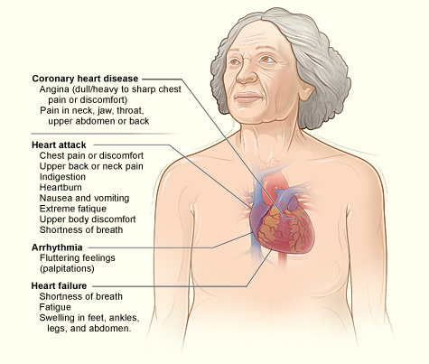
NIH. (n.d.). What is heart disease? Retrieved from website: http://www.nhlbi.nih.gov/health/dci/Diseases/hdw/hdw_all.html
Parisi, Mark. (n.d.). Menopause Cartoon # 1996-11-10. Retrieved from website: http://offthemark.com/search-results/key/menopause/